Personalised Virtual Brain Twins: the Future of Neuroscience and Medicine
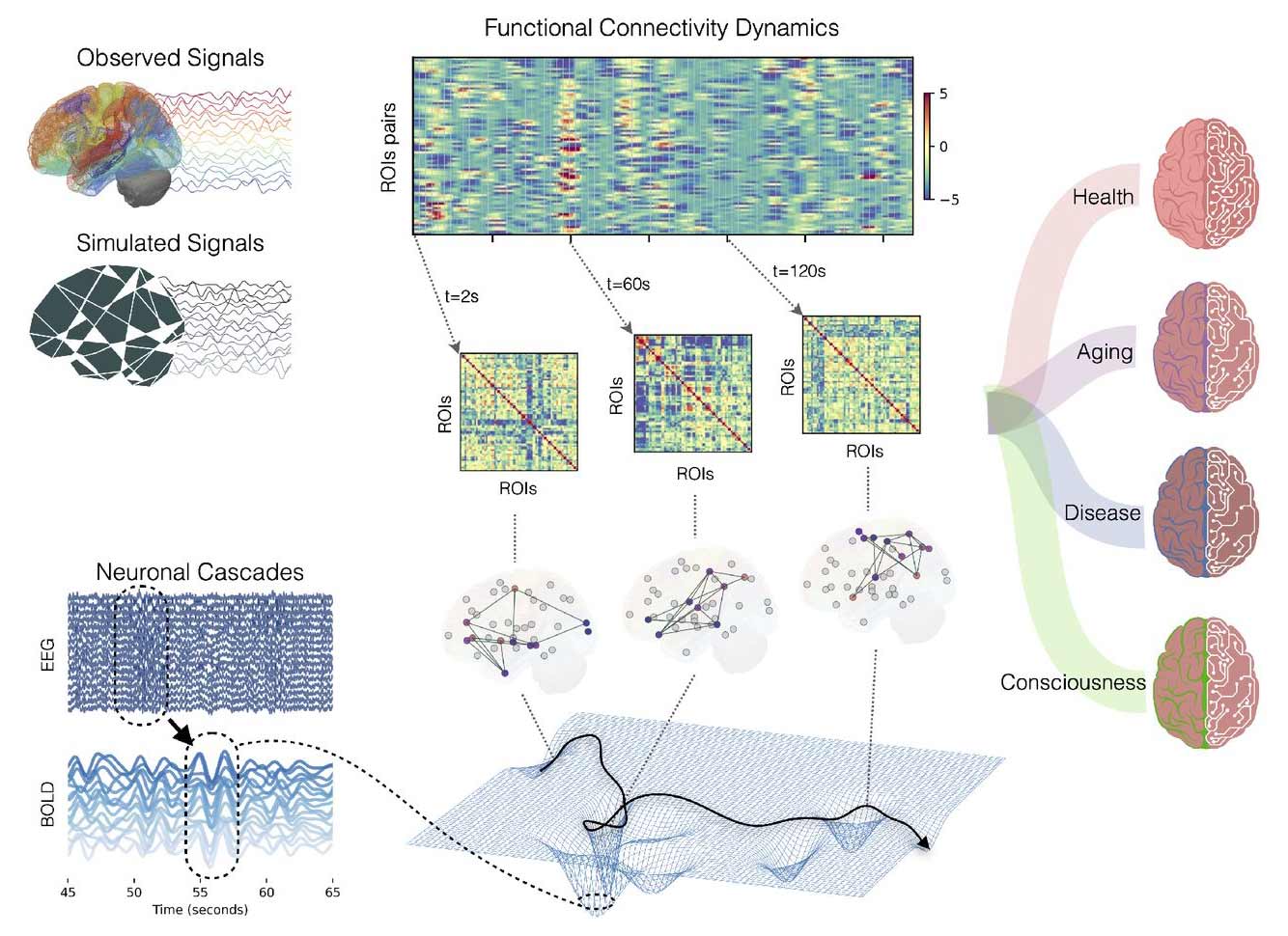
VBT of resting- and altered-states.
Most current medical decisions consider patients as part of a general population, applying treatments based on averages. But no two brains are the same. The use of a Virtual Brain Twin (VBT) could change this approach by creating a personalised digital replica of an individual’s brain network, opening new frontiers in neuroscience and medicine.
What is a Virtual Brain Twin?
A Virtual Brain Twin (VBT) combines a person’s structural brain data (from MRI or diffusion imaging) with functional data which measure brain activity (such as EEG, MEG, or fMRI) to build a computational model of their brain’s network. This network model simulates how different brain regions are connected and communicate through neural pathways.
How is a VBT built?
This article covers the core conceptual, mathematical, technical and clinical aspects of how VBTs are developed (1) - from scanning and modelling the brain anatomy to running simulations and Bayesian inference (2). The resulting VBT can then be used to test “what-if” scenarios in a safe, virtual environment - before applying treatments or interventions in the real world, i.e. on the patient’s brain.
Applications in research and medicine
Since VBTs are grounded in biological principles, they can help clinicians see how microscopic changes at the cellular level affect whole-brain dynamics, through brain imaging and simulations. These models enable predictions on how an individual might respond to specific interventions and can help clinicians in managing neurological and psychiatric conditions, in predicting drug side-effects, in tailoring non-invasive brain stimulation, and in the customisation of rehabilitation programmes.
Researchers also demonstrate how VBTs could be applied to:
- Epilepsy – The Virtual Epileptic Patient model helps identify seizure origins and evaluate precise tailored surgical strategies and electrical stimulation methods for drug-resistant cases.
- Multiple sclerosis – Modelling how lesions disrupt brain connectivity can inform rehabilitation and therapy planning.
- Healthy ageing and Alzheimer’s disease – Tracking changes in network dynamics over time may improve early diagnosis.
- Resting-state brain activity – Understanding deviations from normal patterns/functions aids in detecting neurological disorders.
In the future, VBTs could help with other brain conditions like Parkinson’s disease, expand the understanding of cognitive functions, guide the development of brain-computer interfaces, and even contribute to consciousness research.
A step towards personalised medicine
By offering interpretable predictions and insights, VBTs allow clinicians to tailor treatments to the individual, marking a shift from “one-size-fits-all” medicine towards precision healthcare - where each patient’s brain is understood, modelled, and treated in its own unique way.
For deeper insights into the VBT's origin, use and promises read the full publication ‘’Principles and Operation of Virtual Brain Twins.’’
Original publication
Meysam Hashemi, Damien Depannemaecker, Marisa Saggio, Paul Triebkorn, Giovanni Rabuffo, Jan Fousek, Abolfazl Ziaeemehr, Viktor Sip, Anastasios Athanasiadis, Martin Breyton, Marmaduke Woodman, Huifang Wang, Spase Petkoski, Pierpaolo Sorrentino, Viktor Jirsa, ‘’Principles and Operation of Virtual Brain Twins'', IEEE Reviews in Biomedical Engineering (2025). https://ieeexplore.ieee.org/document/10972118/
Notes
1. The creation of a VBT involves three key steps:
- a. Structural mapping – Brain scans are processed to identify regions (nodes) and the white-matter connections between them (the connectome).
- b. Model integration – Each node is assigned a mathematical model (a Neural Mass Model) representing the average activity of large groups of neurons, while the connectome defines how signals travel between regions.
- c. Personalisation through data – Functional brain recordings are used to fine-tune the model using statistical inference, adapting it to the individual’s physiology and condition.
2. Bayesian inference is the process of updating the probability of a hypothesis—originally based on incomplete information and prior beliefs—when new evidence becomes available.
Glossary
- MRI – Magnetic Resonance Imaging, a method for visualising brain structure.
- EEG / MEG / fMRI – Techniques for measuring brain activity.
- Connectome – Comprehensive map of brain connections.
- Neural Mass Model – Mathematical representation of the activity of a large group of neurons.